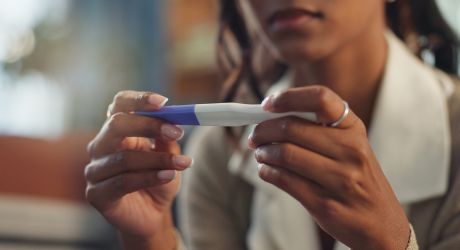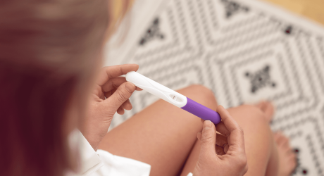
The first ultrasound after fertility treatment is one of the most anticipated moments following a positive pregnancy test.
Although the pregnancy test confirms the pregnancy, it is common for uncertainty to continue until an ultrasound confirms that the pregnancy is progressing correctly.
This first examination makes it possible to confirm the location of the pregnancy and assess whether the pregnancy is developing as expected.
When is the first ultrasound performed after in vitro fertilisation (IVF) or egg donation?
In general, the first ultrasound after IVF or egg donation is performed approximately two weeks after a positive pregnancy test.
At this early stage of pregnancy, the fetal heartbeat may not yet be visible, but the ultrasound already provides very important information about the progression of the pregnancy.
This examination can help to:
- Rule out an ectopic pregnancy (located outside the uterus).
- Determine whether it is a singleton or multiple pregnancy.
- Assess whether early embryonic development is as expected.
What can be seen in the first ultrasound?
During the first weeks of pregnancy, different embryonic structures may be observed.
Gestational sac
This is the first structure visible on ultrasound. It appears as a small rounded structure within the uterus, confirming that the pregnancy has implanted correctly.
Yolk sac
The yolk sac appears at very early stages of embryonic development and is usually seen inside the gestational sac. Its presence is one of the earliest signs of a normally developing pregnancy.
Embryonic pole
The embryonic pole represents the earliest visible stage of embryonic development. In the first weeks, it may be seen as a small structure next to the yolk sac, although in some cases it may not yet be visible at week 6.
Embryonic heartbeat
The embryonic heartbeat is usually visible from week 6 of gestation. However, not seeing it on a very early ultrasound does not necessarily mean there is a problem. As the pregnancy progresses, the heartbeat becomes easier to detect and the heart rate increases progressively during the first weeks.
Pregnancy monitoring after fertility treatment
Although several follow-up ultrasounds are usually performed during pregnancy, the number and frequency will depend on the characteristics of each pregnancy and the presence or absence of risk factors.
At Barcelona IVF , monitoring after fertility treatment is tailored to each patient, supporting each patient throughout the entire process.